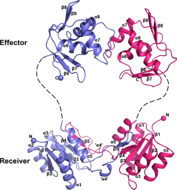
Figure 1.

Ribbon representation of the BaeR dimer. One protomer is shown in light blue and the other in magenta. The linker region is missing from the final structure but for visualization purposes we have drawn it as dotted line (Supporting Information Fig. S2). The complete receiver domain is formed from a domain swap between the β-core domain and α4-β5. An interactive view is available in the electronic version of the article. [Color figure can be viewed in the online issue, which is available at http://wileyonlinelibrary.com.]
