Figure 1.
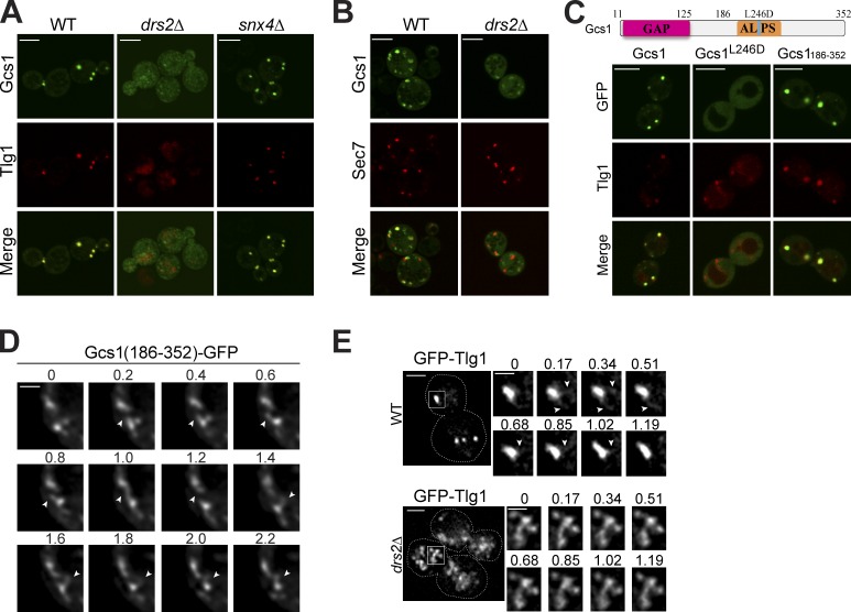
Drs2 flippase activity and the ALPS motif are required for Gcs1 TGN/EE localization. (A) Gcs1-GFP localization in wild-type (WT), drs2Δ, and snx4Δ cells relative to mCherry-Tlg1. (B) Gcs1-GFP localization in WT and drs2Δ cells relative to Sec7-DsRed. (C) GFP-tagged Gcs1, Gcs1L246D, and Gcs1(186–352) expressed in WT cells were imaged relative to mCherry-Tlg1. (D) Time-lapse images of Gcs1(186–352)-GFP decorating small diameter tubules (arrows) extending from TGN/EE. Time is indicated in seconds. (E) Membrane dynamics of TGN/EE in WT and drs2Δ cells. GFP-Tlg1 was imaged for several seconds (Videos 1 and 2), and time-lapse series for the boxed TGN/EE membranes are shown. Time is indicated in seconds. Bars: (A–C) 5 µm; (D) 1 µm; (E, main panels) 2 µm; (E, time lapse) 1 µm.

