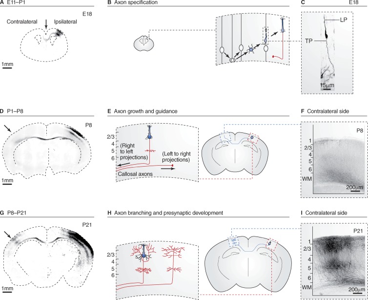
Figure 1.
Axon specification, growth, and branching during mouse cortical development. Three stages of the development of callosal axons of cortical pyramidal neurons from the superficial layers 2/3 of the somatosensory cortex in the mouse visualized using long-term in utero cortical electroporation. For this class of model axons, development can be divided in three main stages: (1) neurogenesis and axon specification, occurring mostly at embryonic ages (A–C); (2) axon growth/guidance during the first postnatal week (D–F); and (3) axon branching and synapse formation until approximately the end of the third postnatal week (G–I). A, D, and G show coronal sections of mouse cortex at the indicated ages after in utero cortical electroporation of a GFP-coding plasmid at E15.5 in superficial neuron precursors in one brain hemisphere only (GFP signal in inverted color, dotted line indicates the limits of the brain). B, E, and H are a schematic representation of the main morphological changes observed in callosally projecting axons (red) at the corresponding ages. C shows the typical bipolar morphology of a migrating neuron emitting a trailing process (TP) and a leading process (LP) that will ultimately become the axon and dendrite, respectively. F and I show typical axon projections of layer 2/3 neurons located in the primary somatosensory area at P8 and P21, respectively. Neurons and axons in C, F, and I are visualized by GFP expression (inverted color). Image in C is modified from Barnes et al. (2007) with permission from Elsevier. Images in D, F, G, and I are reprinted from Courchet et al. (2013) with permission from Elsevier.

