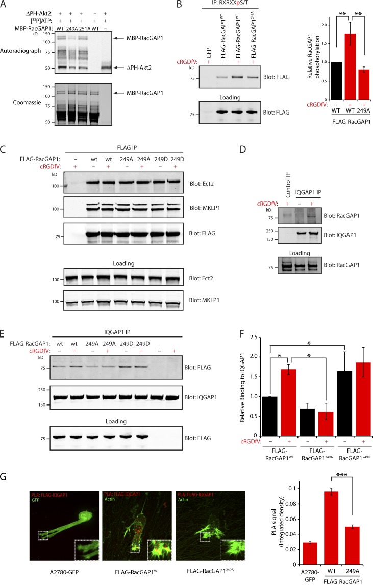
Figure 3.
RacGAP1 phosphorylation on T249 promotes association with IQGAP1. (A) Purified MBP-RacGAP1 and mutants were subjected to in vitro phosphorylation and incorporation of 33P measured as in Fig. 1 C. (B) PKB/Akt substrates were immunoprecipitated from lysates of A2780 cells stably expressing GFP, FLAG-RacGAP1, or FLAG-RacGAP1249A as in Fig. 1 D. (C) Lysates of A2780 cells stably expressing GFP(−), FLAG-RacGAP1, FLAG-RacGAP1249A, or FLAG-RacGAP1249D were subjected to IP using FLAG antibodies. IPs were analyzed by SDS-PAGE and Western blotting for FLAG, Ect2, and MKLP1. (D) Lysates of A2780 cells were subjected to IP using rabbit IQGAP1 antibodies or an isotype-matched control. IPs were analyzed by SDS-PAGE and Western blotting for RacGAP1 and IQGAP1. (E) Cells as in C were lysed, and IP was performed using rabbit IQGAP1 antibodies. IPs were analyzed by SDS-PAGE and Western blotting for FLAG and IQGAP1. (F) Western blots from IPs as in E were quantified using the Odyssey system. (G) Cells stably expressing GFP, FLAG-RacGAP1WT, or FLAG-RacGAP1249A on CDM were treated with cRGDfV for 1 h and fixed and stained with antibodies against FLAG and IQGAP1 before performing PLA. PLA signal was quantified by measuring the integrated density within the whole cell using ImageJ (n > 60/condition). Zoomed insets correspond to areas indicated by dotted ROIs. Bars, 20 µm. Data represent means ± SEM from at least three independent experiments. *, P < 0.05; **, P < 0.01; ***, P < 0.001.

