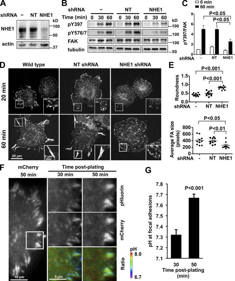
Figure 1.
NHE1 shRNA and decreased pHi inhibit FAK-pY397 and cell spreading. (A) Decreased NHE1 expression in MEFs infected with lentivirus encoding NHE1 shRNA compared with uninfected (−) cells and cells infected with nontargeting (NT) shRNA, determined by immunoblotting cell lysates. (B) FAK-pY397 and -pY576 were attenuated with NHE1 shRNA compared with control (−) and NT shRNA, as indicated by immunoblotting lysates of cells in suspension (0 min) and plated on fibronectin (60 min). (C) Abundance of FAK-pY397 relative to total FAK at the indicated times on fibronectin. Data are means ± SEM of three cell preparations. (D and E) In MEFs plated on fibronectin for 20 and 60 min and immunolabeled with anti-paxillin antibodies, NHE1 shRNA impairs cell spreading, indicated by roundness index, and focal adhesion size compared with control (−) and NT shRNA. Insets are enlarged images of boxed areas of representative focal adhesions. (F) Time-dependent increase in pH at focal adhesions with plating on fibronectin determined by TIRF ratiometric imaging of pH-sensitive pHluorin and pH-insensitive mCherry fused to paxillin. 10-µm × 10-µm images of cells (marked at left panel) at 30 and 50 min are shown. (G) Mean pH ± SEM of five focal adhesions at 30 and 50 min are representative of cells from three independent preparations.

