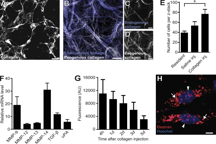
Figure 1.
An assay to visualize intracellular collagen degradation in situ. (A–D) Microscopic appearance of collagen fibers formed after incubation of Alexa Fluor 647–labeled acid-extracted rat tail tendon type I collagen at neutral pH in vitro (A) or formed in vivo 24 h after injection into the dermis of live mice (B–D). Dermal-injected fluorescently labeled rat tail collagen fibrils form collagen fiber networks (white fibers in B) that may be associated with existing collagen/elastin scaffolds (blue fibers in B and white fibers in C, visualized by confocal microscopy) or may form ab initio (compare C and D). (E–G) Dermal collagen injection disrupts tissue homeostasis and generates a matrix catabolic environment. (E) Total cell counts in noninjected (left bar), vehicle-injected (middle bar), or collagen-injected (inj.; right bar) dermis, as determined by the counting of nuclei in four to six serial z stacks from the injection field. n = 3 for each treatment. *, P < 0.05, Student’s t test, two tailed. (F) Quantitative PCR analysis of mRNA levels of six genes associated with extracellular matrix remodeling in the dermis of collagen-injected mice. Data are expressed as fold increase relative to noninjected mice. n = 3 for noninjected and for collagen-injected mice. uPA, urokinase-type plasminogen activator. (G) Total fluorescence extracted at the indicated time points from skin of mice injected with Alexa Fluor 488–conjugated collagen. n = 4 for each time point. AU, arbitrary unit. (H) Visualization of the lysosomal compartment (red; examples with arrows) and nuclei (blue; arrowheads) of cells in dermal injection fields by local injection with Texas red 10-kD dextran and systemic injection of Hoechst dye, respectively, 24 and 4–6 h before imaging. Data are shown as means ± SD. Bars: (A–D) 10 µm; (H) 5 µm.

