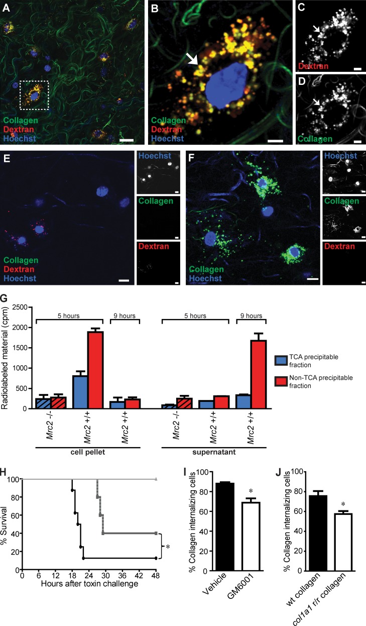
Figure 2.
Cellular uptake and lysosomal routing of collagen in vivo. (A) Representative image of mouse dermis 24 h after injection shows vigorous cellular uptake of collagen, as revealed by multiple cells presenting with fluorescently labeled collagen located within perinuclear endocytic vesicles. (B–D) High magnification of a single collagen-uptaking cell (box in A), illustrating the frequent colocalization of collagen and Texas red dextran in perinuclear vesicles (yellow; examples with arrows), indicative of lysosomal routing of endocytosed collagen. (E) Representative image of mouse dermis distant to the site of collagen and dextran injection. (F) Representative image of mouse dermis 24 h after injection of fluorescently labeled collagen. (G) Primary fibroblasts from wild-type or littermate uPARAP-deficient mice (hatched bars) were allowed to internalize 125I-labeled collagen for 5 h, after which the cells were trypsinized, washed, and reseeded. At 5 and 9 h after collagen exposure (0 and 4 h after reseeding), the cells (left) and the supernatant (right) were separated, and radioactivity in the TCA-precipitable fraction and the non-TCA–precipitable fraction was measured. Error bars indicate SDs. (H and I) Effect of systemic metalloproteinase inhibition on intracellular collagen degradation. (H) GM6001 treatment of mice inhibits MMP activity in vivo. Mice were treated with GM6001 or vehicle 24 and 0.5 h before i.p. injection with either the MMP-activated toxin PrAg-L1 or the nonactivatable control toxin PrAg-U7 (circles, vehicle + PrAg-L1 [n = 8]; squares, GM6001 + PrAg-L1 [n = 5]; triangles, vehicle + PrAg-U7 [n = 5]). *, P = 0.041, log-rank test. (I) Percentage of collagen-internalizing cells in the dermis of vehicle-treated or GM6001-treated mice. n = 3 for each group of collagen-injected mice treated with vehicle or with GM6001. *, P < 0.05, Student’s t test, two tailed. The quantitative data in this and the following figures are shown as mean ± SD and were obtained by counting 40–120 cells per z stack from four to six serial z stacks per mouse. (J) Percentage of collagen-internalizing cells in the dermis of mice injected with wild-type (wt) collagen or collagenase-resistant (Col1a1r/r) collagen. n = 4 for each group of collagen-injected mice. *, P < 0.01, Student’s t test, two tailed. Bars: (A) 50 µm; (B–D) 10 µm; (E and F) 15 µm.

