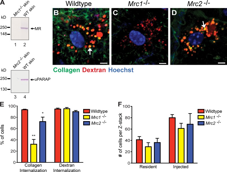
Figure 5.
MR and uPARAP facilitate intracellular collagen degradation in vivo. (A) Western blot of MR (lanes 1 and 2) and uPARAP (lanes 3 and 4) in protein extracts from dermis of MR-deficient (Mrc1−/−; lane 1) and a wild-type (WT) littermate (lane 2) or uPARAP-deficient (Mrc2−/−; lane 3) and a wild-type littermate (lane 4). Arrows on the right show the positions of MR and uPARAP. Positions of molecular mass markers (kilodaltons) are indicated on the left. Specificity of MR and uPARAP detection is shown by the absence of immunoreactivity in Mrc1−/− and Mrc2−/− mice, respectively. (B–D) Representative examples of collagen uptake in cells from the dermis of wild-type (B), Mrc1−/− (C), and Mrc2−/− (D) mice 24 h after collagen injection. Bars, 10 µm. Arrows in B and D show examples of collagen in lysosomal vesicles. (E) Percentage of cells in collagen- and dextran-injected wild-type (n = 5), Mrc1−/− (n = 3), and Mrc2−/− (n = 5) dermis that internalize collagen or dextran. *, P < 0.05; and **, P < 0.01 relative to collagen-internalizing cells in wild-type mice (Student’s t test, two tailed). (F) Enumeration of cells present in noninjected or collagen-injected dermis of wild-type (n = 3), Mrc1−/− (n = 3), and Mrc2−/− (n = 3) mice. Error bars show means ± SD.

