
Figure 1.
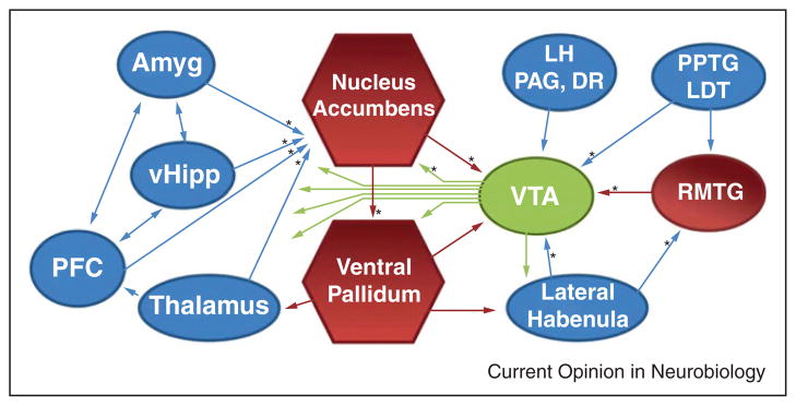
Schematic of the principal brain regions that innervate the VTA and NAc. Red indicates inhibitory structures and blue indicates excitatory structures. Pathways that have been examined with optogenetic techniques are indicated with an *. Amyg, amygdala; vHipp, ventral hippocampus; LH, lateral hypothalamus; PAG, periaqueductal gray; DR, dorsal raphe; PPTG/LDT, pedunculopontine and laterodorsal tegmentum; RMTG, rostromedial tegmental nucleus.