Figure 5.
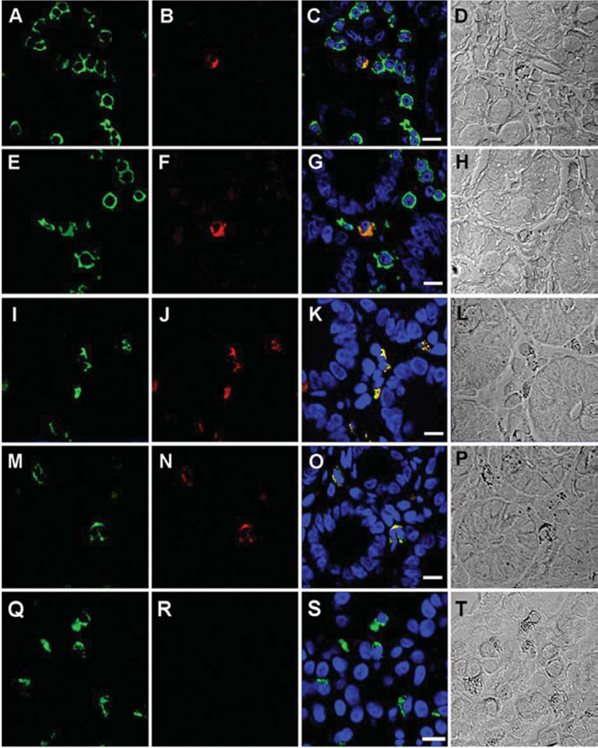
Phenotype of duodenal-associated cells immunoreactive to rat monoclonal antibody to SFFVgp52 Env. A: Expression of CD45 (FITC) in a duodenal biopsy of an ME case. B: Immunoreactivity of rhodamineconjugated monoclonal rat antibody to SFFVgp52 Env in duodenal biopsy of an ME case. C: Merged images A and B, (Bar represents 20 µm). D: Morphology of the immunoreactive cells. E: Expression of CD303 (FITC) in a duodenal biopsy of an ME case. F: Immunoreactivity of rhodamine-conjugated monoclonal rat antibody to SFFVgp52 Env in a duodenal biopsy of an ME case. G: Merged images E and F (Bar represents 20 µm). H: Morphology of the immunoreactive cells. I: Expression of CD86 (FITC) in duodenal a biopsy of an ME case. J: Immunoreactivity of a rhodamine-conjugated monoclonal rat antibody to SFFV-gp-52 Env in a duodenal biopsy of an ME case. K: Merged images I and J, (Bar represents 20 µm). L: Morphology of the immunoreactive cells. M: Expression of HLADR (FITC) in a duodenal biopsy of an ME case. N: Immunoreactivity of rhodamine-conjugated monoclonal rat antibody to SFFV-gp-52 Env in duodenal biopsy of an ME case. O: Merged images M and N, (Bar represents 20 µm). P: Morphology of the immunoreactive cells. Q: Expression of CD303 (FITC) in a duodenal biopsy of an ME case. R: Immunoreactivity of APC-conjugated mouse IgG1 isotype control in duodenal biopsy of an ME case. S: Merged images Q and R (Bar represents 20 µm). T: Morphology of the immunoreactive cells. TOPO3 was used to a indicate nucleus localization in images C, G, K, O and S.

