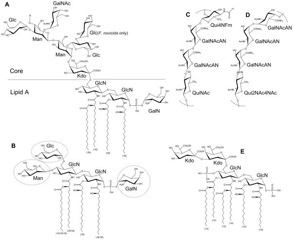
Figure 1. Structure of Francisella LPS.
Major Francisella lipid A molecule with core oligosaccharide (A). The length of each fatty acyl chain is indicated in parenthesis. Chain length variation (16/18) indicates temperature regulation. Less abundant lipid A variants may have various modifications in sugar composition as shown in dashed circles (B). The structures of F. tularensis O-antigen (C), and F. novicida O-antigen (D) are shown. For comparison, E. coli Kdo2-lipid A is also shown (E). R = O-antigen.

