
Figure 6.
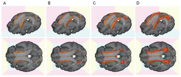
Three-dimensional white matter hyperintensity (WMH) frequency maps for each cognitive group. (A) First quartile Mini-Mental State Examination (MMSE) 29 to 30; (B) second quartile, MMSE 28; (C) third quartile, MMSE 25 to 27; (D) fourth quartile MMSE 0 to 24. Orange area indicates voxels containing WMH with a frequency of 10% or higher.