Figure 3.
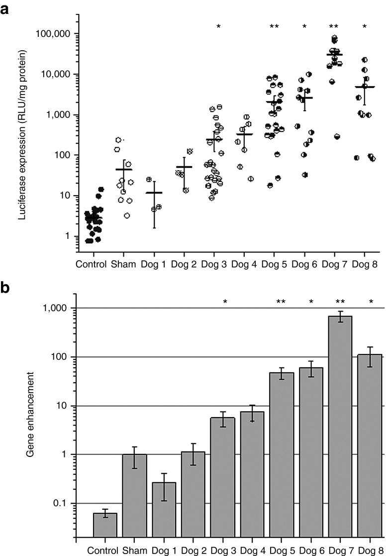
Luciferase gene expression following ultrasound-targeted microbubble destruction (UTMD)-mediated gene delivery into the dog livers. (a) Four milligrams of pGL4 plasmid and 1.5–3 ml of Definity MBs were injected via the portal vein (PV) or the segmental PV branch with simultaneous exposure of the target liver lobe to therapeutic US (tUS) (1.1 MHz frequency, 20 cycle pulses, 13.9–50 Hz pulse repetition frequency, and 2.0–3.3 MPa peak negative pressures (PNPs)) for 2–4 minutes using the small diameter transducer (H158) for dog experiments 1–3, or the large diameter transducer (H105) for dog experiments 4–8. A sham-treated dog received an equivalent pGL4/MB dose, but was not exposed to tUS (or 0 MPa PNP tUS exposure). Treated and untreated control lobes were harvested after 24 hours. Each lobe was sectioned, and the representative sections (shown as data points) were processed and assayed for luciferase activity. The average luciferase expression for each treated lobe is shown as horizontal lines, and the error bars indicate SEM (n = 3–15). *P < 0.05, **P < 0.005. (b) The average luciferase expression from each treated dog was normalized to the average luciferase expression of the sham-treated dog to determine the overall gene enhancement using modified surgical techniques and UTMD methods. Data showed stepwise improvement in gene enhancement with each dog experiment as occlusion strategies, proper MB injection site, and better tUS transducers and instrumentation were used. Error bars indicate SEM (n = 3–15). *P < 0.05, **P < 0.005. RLU, relative light unit.

