
Figure 6.
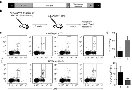
Expression of Tregitopes in vivo results in modulation of capsid-specific IFN-γ responses. (a) Diagram of the transgene expression cassette used to co-express AAV capsid antigen (AAV2VP1) and Tregitope or Scramble peptides. ITR, inverted terminal repeats; CMV, cytomegalovirus enhancer/promoter; RKR, PACE/Furin cleavage sequence; pA, poly-A signal. (b) Experimental design. At day 0, C57BL/6 mice received the Tregitope vector or the Scramble control intramuscularly, 2 × 10¹¹ vg/mouse; six weeks later, animals were immunized with an adenoviral vector expressing the AAV2 VP1 protein injected intramuscularly, 1 × 10¹¹ vp/mouse. Nine days after Ad immunization animals were sacrificed. (c) Intracellular cytokine staining for IFN-γ after AAV peptide restimulation of splenocytes isolated from animals 9 days after adenoviral challenge. After live/dead exclusion, cells were gated on lymphocytes, and CD8+ T cells were then analyzed for IFN-γ positivity. (d) Histogram plot of the frequency of CD8+IFN-γ+ T cells shown in panels (c) in animals receiving the AAV-Tregitope (T) or AAV-Scramble (S) vectors, the y-axis represents the percent of total lymphocytes that are CD8+IFN-γ+; *P = 0.0428. (e) Frequency of CD4+CD25hi cells that are FoxP3+ in splenocytes of animals receiving the AAV-Tregitope (T) or AAV-Scramble (S) vectors; after live/dead exclusion, cells were gated on lymphocytes, CD4+ T cells, and CD25hi cells were analyzed for FoxP3 expression; the y-axis represents the percent of CD4+ T cells that are CD25hiFoxP3+; *P = 0.0256. Shown are individual animals from one of three independent experiments. Error bars represent SEM.