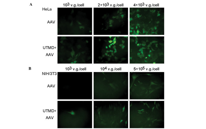
Figure 3.
Fluorescence microscopy of enhanced green fluorescent protein (EGFP) expression. (A) Fluorescence microscopy of HeLa cells 48 h following infection with recombinant adeno-associated virus serotype 2 (rAAV2)-EGFP with three different multiplicities of infection (MOIs). The cells in the AAV group were infected with rAAV2-EGFP alone. The cells in the ultrasound-targeted microbubble destruction (UTMD)+AAV group were infected with rAAV2-EGFP and treated with optimized UTMD. (B) Fluorescence microscopy of NIH/3T3 cells 48 h following infection with rAAV2-EGFP with three different MOIs. v.g., vector genome.

