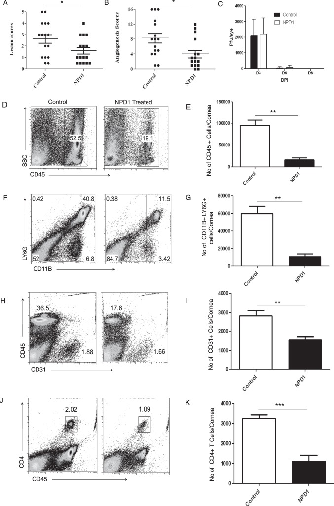
Figure 1.
Effect of prophylactic treatment with NPD1 on SK severity and cellular infiltration. C57BL/6 mice infected with 2 × 10⁴ PFU of HSV-1 RE were given NPD1 topically twice daily starting from 1 day before infection until day 10 pi. The disease severity was examined on different days after infection. (A, B) SK lesion severity and angiogenesis scores at day 15 pi are shown. The level of significance was determined by Student's t-test (unpaired, n = 16–18 mice/group as indicated in the scatter plots). (C) At the indicated time points, eyes of HSV-infected mice were swabbed with a sterile swab and assayed for infectious virus by standard plaque assay. The level of significance was determined by Student's t-test (unpaired). Error bars represent mean ± SEM (n = 10 eyes). (D–K) The immune parameters were analyzed at the termination of the experiment (day 15 pi). Representative histograms show percentage of (D) leukocytes (CD45+), (F) neutrophils (CD11B+Ly6G+), (H) CD31+ cells, and (J) CD4+ T cells in the inflamed cornea of control and NPD1-treated animals at day 15 pi. Average numbers of (E) CD45+ cells, (G) CD11B+Ly6G+, (I) CD31+ cells, and (K) CD4+ T cells per cornea at indicated time point are shown. The level of significance was determined by Student's t-test (unpaired). Error bars represent mean ± SEM, n = 4 corneas per group. Experiments were repeated at least two times. *P ≤ 0.05. **P ≤ 0.01. ***P ≤ 0.001.

