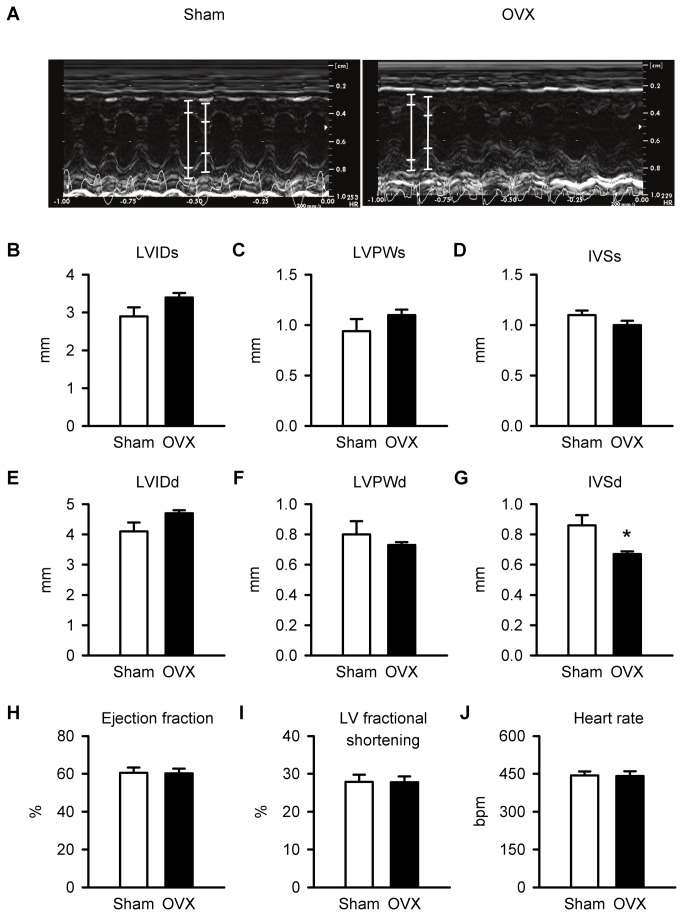
Figure 2. In vivo ventricular structure, function and heart rate were similar in sham and OVX mice.
A. M-mode images of cardiac function from sham and OVX mice. The white lines in each image denote the lumen edges in systole and diastole. B,C,D. Left ventricular internal diameter in systole (LVIDs), left ventricular posterior wall thickness in systole (LVPWs) and interventricular septal thickness in systole (IVSs) were similar in sham and OVX hearts. E,F. Left ventricular internal diameter in diastole (LVIDd) and left ventricular posterior wall thickness in diastole (LVPWd) were unaffected by OVX. G. By contrast, interventricular septal thickness in diastole (IVSd) was reduced significantly by OVX. H,I,J. Mean values for ejection fraction, left ventricular fractional shortening and heart rate were identical in sham and OVX mice (n=6 sham and 6 OVX mice; *p<0.05; t-test).

