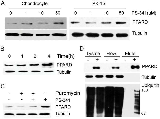
Figure 4. sPPARD turnover is controlled by ubiquitin-proteasome system.
(A) Chondrocyte and PK-15 cells were treated with PS341 at 4 different concentrations for 4 h, and sPPARD levels were detected by Western blot. (B) PK-15 cells were treated with 10 µM PS341 and sPPARD levels were detected by Western blot at four time points after the treatment. (C) PK-15 cells were treated with puromycin, puromycin and PS341, or PS341 alone for 4 hours and sPPARD levels were detected by Western blot. (D) HEK293T cells were transfected with HA-ub vector along with pcDNA4A-His or –sPPARD expression vectors and after 24 hours were incubated with 10 µM PS341 for 4 hours. His-tagged sPPARD was pulled down with nickel affinity gel under denaturing conditions. Expression levels of sPPARD and tubulin in cell lysates are shown below. An arrow and a bar indicate mono-ubiquitinated and poly-ubiquitinated sPPARD, respectively. sPPARD was detected in cell lysate, flow-through, and eluate fractions.

