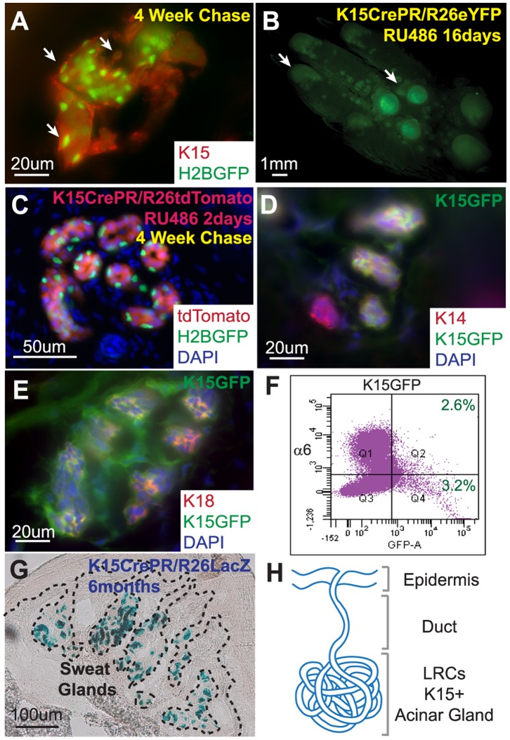
Figure 3. Sweat gland LRCs express Keratin 15 and contribute long term to the acinar SG structure.
(A) K15 staining of sweat gland LRCs indicate positive K15 expression. (B) Fluorescent photo of K15CrePR/R26eYFPRU palm containing YFP positive sweat glands after long term YFP activation. (C) Section of K15CrePR/R26tdTomRU crossed onto K5TetOff/TreH2BGFP sweat glands after 4 weeks of chase with doxycycline followed by 2 days of RU treatment. (D) K14 basal layer staining co-localizes with GFP expression in K15-GFP transgenic sweat glands. (E) K18 lumenal marker staining co-localizes with K15-GFP expression in sweat glands. (F) FACS analysis of K15-GFP sweat glands demonstrates that approximately half of the K15-GFP positive cells are localized to the basal layer expressing α6 integrin. (G) Histology of X-Gal-treated K15CrePR/R26LacZ transgenic mice, blue stain indicates transgene expression for more than 6 months after RU activation in sweat glands. (H) K15 expression co-localizes with LRCs in the acinar sweat gland region.

