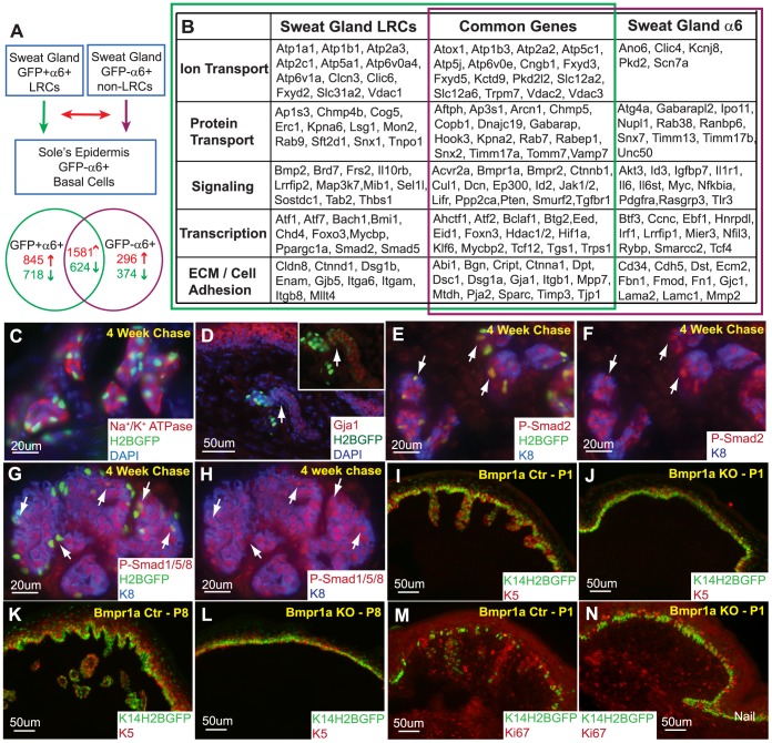
Figure 5. Molecular characteristics of sweat gland LRCs define BMP signaling as a requirement for SG formation.
(A) GFP+/α6+ sweat gland LRCs and GFP−/α6+ sweat gland non-LRCs basal cells were compared to the basal layer of the sole’s epidermis. The resulting gene expression profiles of these sweat gland LRCs and basal cells were then compared to each other. (B) Gene expression profiles consistently found in two independent microarray analyses from independent biological samples were categorized into ion and protein transport, signaling, transcription, extracellular matrix (ECM) and cell adhesion based on function. (C) Sodium Potassium ATPases were confirmed to be expressed in the sweat glands. (D) Gja1 is confirmed to be expressed in SG LRCs (arrows) as well as non-LRCs. (E) Phospho-Smad2 co-localizes with SG LRCs, arrows. (F) Corresponding phospho-Smad2 and K8 channels. Arrows indicate LRCs marked in panel “E”. (G) Positive phospho-smad1/5/8 staining indicates active BMP signaling in sweat glands. (H) Corresponding phospho-Smad1/5/8 and K8 channels with arrows indicating co-localization with some LRCs. (I) Downgrowth of sweat glands is observed in P1 control paws but is absent (J) in Bmpr1a/K14Cre/K14-H2BGFP KO paws. (K) Similarly, more developed sweat glands are observed in P8 control paws but are still absent (L) in KO mice. (M) The basal layer of the epidermis as well as the sweat glands is proliferative at P1. (N) Although sweat glands are absent in Bmpr1a/K14Cre/K14-H2BGFP KO paws, the epidermal basal layer is still capable of division.

