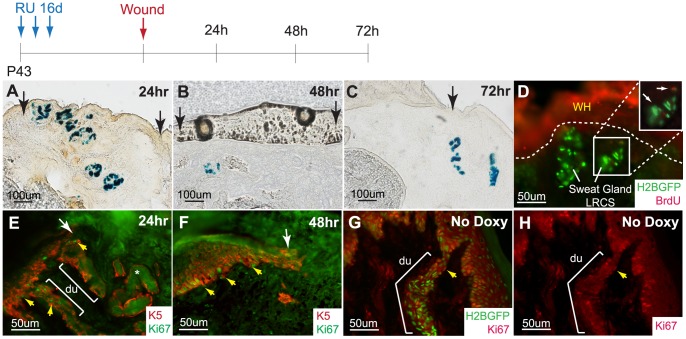
Figure 6. Acinar sweat gland cells do not contribute to the epidermis during typical wound healing.
(A) K15CrePR/R26LacZRU marked sweat gland cells do not contribute to the epidermis at 24 h, (B) 48 h, and (C) 72 h after wounding. (D) BrdU pulse shows that a few SG cells are activated upon injury (inset, arrows) while most SG LRCs remain quiescent. (E) Ki67 staining confirms that the acinar sweat gland region is quiescent while the SG duct and epidermal basal layer is proliferative at 24 h and (F) 48 h. (G) Under normal homeostasis, cells of the SG duct and epidermal basal layer are active in the cell cycle. (H) Corresponding single Ki67 (red) channel. Abbreviations: du, sweat ducts.

