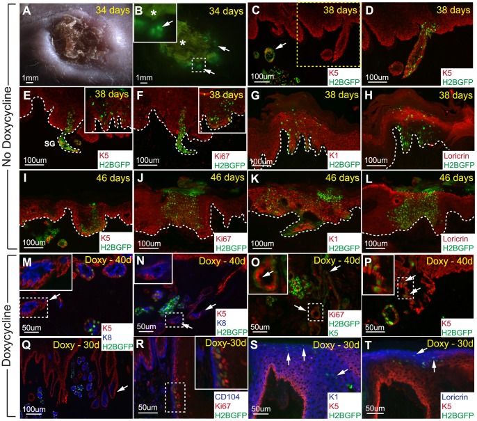
Figure 7. Sweat gland LRCs can trans-differentiate into the epidermis under prolonged isolated wound healing conditions.
(A) DIC photo of the back skin area containing transplanted H2BGFP labeled sweat glands at 34 days. (B) Corresponding fluorescent image displaying the presence of H2BGFP positive cells in the grafted area. Arrows indicate regions with H2BGFP positive cells while “*” marks autofluorescence of the wounded area. (C) Biopsy was taken at 38 days where H2BGFP sweat glands were observed in the dermis, arrow. (D) Higher exposure time for the GFP channel on the same section from “C” detected marked cells with lower H2BGFP intensities in the epidermis. (E,I) K5 basal layer staining demonstrating the contribution of sweat gland cells to the newly formed epidermis at 38 and 46 days, respectively. (F,J) These H2BGFP labeled cells are still able to proliferate as indicated by Ki67 staining. (G,K) Sweat gland cells can differentiate into cells of the suprabasal layer marked by K1 at 38 and 46 days, respectively. (H,L) These sweat gland cells can also contribute to the granular layer as marked by loricrin. (M,N) When 4 weeks chased sweat glands are transplanted and kept on doxycycline treatment, the H2BGFP label gets diluted out of some sweat glands (arrows), as confirmed by K5 and K8 staining for sweat glands. (O) Ki67 staining show that these sweat glands lacking visible H2BGFP LRCs (green - nuclear) defined by K5 positive staining (green membrane staining) contains Ki67 positive dividing cells (arrows). (P) H2BGFP sweat gland LRCs can contribute to the newly formed epidermis as indicated by K5 basal layer staining (arrows). (Q) Some sweat glands are connected to the newly formed epidermis lacking visible H2BGFP LRCs. (R) Sweat gland LRCs contributing to the epidermis are proliferative near the CD104 marked epidermal basal layer as marked by Ki67, inset shows magnification of co-localization. (S) Sweat gland LRCs can contribute to the suprabasal layer marked by K1 as well as the (T) granular layer expressing loricrin. White dotted lines mark dermal-epidermal interfaces. Abbreviations: CD104, β4 integrin.

