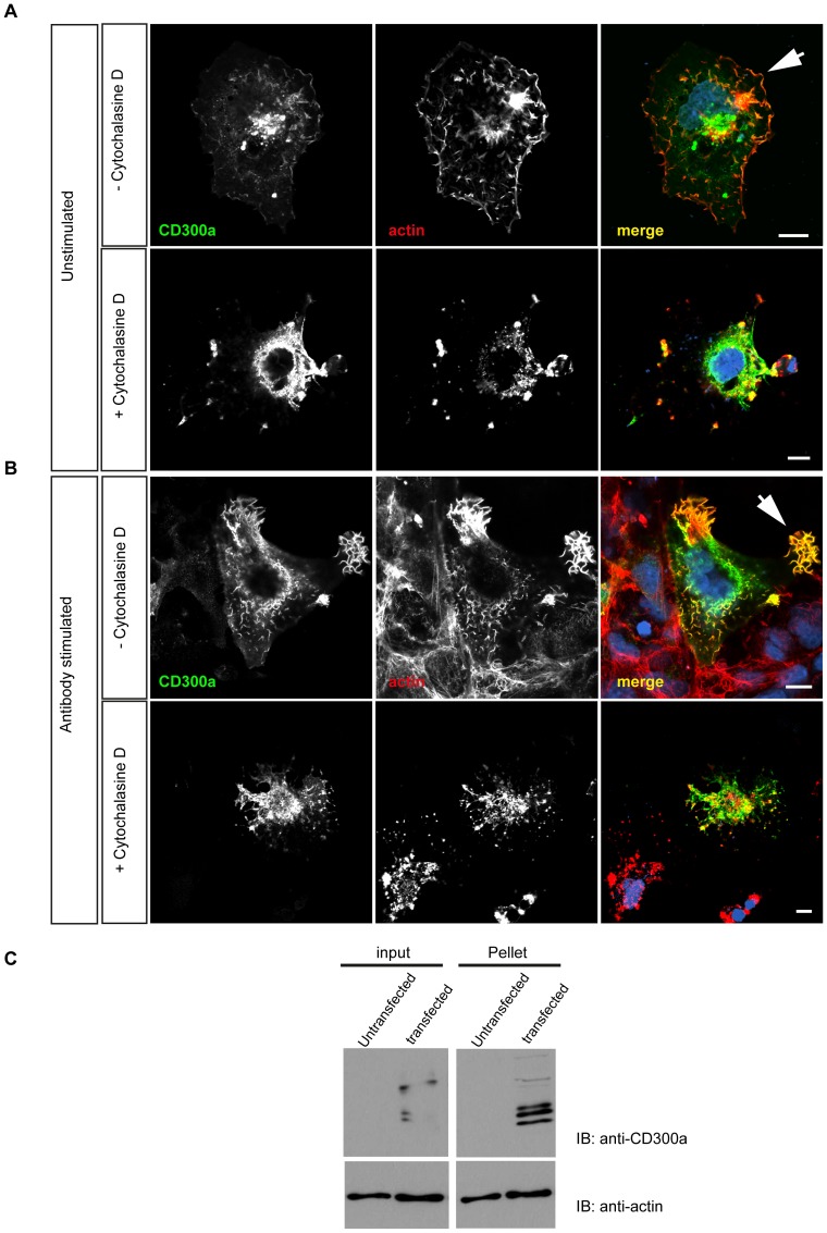
Figure 5. CD300a associates with F-actin in COS-7 cells.
A. COS-7 cells were transfected with a CD300a expression construct and the subcellular localization of CD300a was then revealed by staining with anti-CD300a antibodies. TRITC-phalloidin was used to label the actin cytoskeleton. The bottom row of images shows cells treated with cytochalasin D (4 μM) to disrupt actin filaments. B. COS-7 cells ectopically expressing CD300a were incubated with monoclonal anti-CD300a antibodies to activate cell surface receptors. Cells were then left untreated or treated with 4 μM cytochalasin D followed by fixation and staining with anti-CD300a antibodies and TRITC-phalloidin. Note the localization of CD300a to actin-rich cell protrusions that is increased following the anti-CD300a antibody treatment (arrowhead). Scale bars, 10 μm C. Coprecipitation of CD300a with actin filaments. Triton lysates of control (non-transfected) or CD300a expressing COS-7 cells (transfected) were treated with nocodazole to depolymerize microtubules and then subjected to high speed centrifugation to pellet actin filaments. CD300a protein was detected in the actin pellet fraction suggesting a direct or an indirect association of the protein with F-actin. Input shows protein levels in the cell lysates prior to high speed centrifugation.

