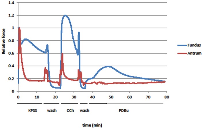
Figure 1. Force (normalized to peak force from KPSS stimulation) generated by stimulating with KPSS, 1 µM CCh, or 1 µM PDBu in antrum (red) and fundus (blue).
Antrum contractile response is not maintained during the stimulation while that in the fundus is. PDBu generated a slow contraction in the fundus that is ∼40% of its peak KPSS force, but caused little to no contraction in the antrum. Deflections in the traces at the start of each wash are from changing the solution in the chambers. These are representative traces from 1 of 6 different animals used for mechanical measurements.

