
Figure 3. Confocal images of PKCα distribution in transverse sections of the longitudinal and circular layer of pig stomach fundus (left) and antrum (right) under relaxed (PSS) or stimulated (3′ CCh or 10 & 30′ PDBu) treatments.
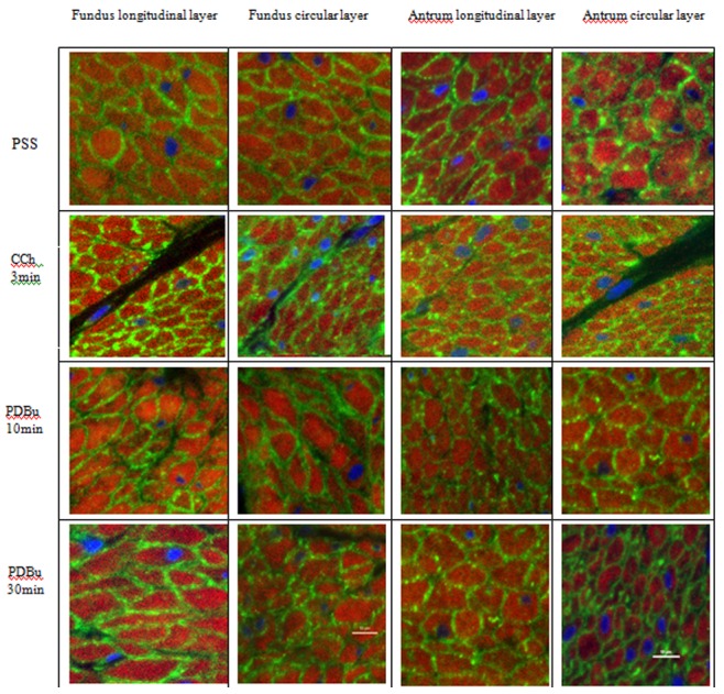
Tissues were immunoreacted for PKCα (green) and counterstained for filamentous actin (phalloidin - red) and nuclei (DAPI - blue). In all conditions, PKCα is located predominantly at the cell periphery near the plasma membrane. Scale bar −10 µm.