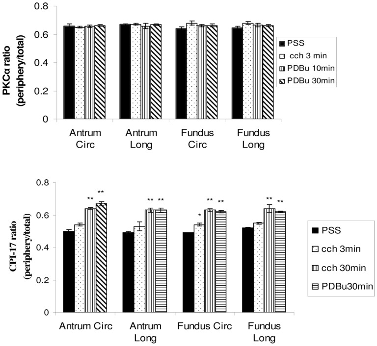
Figure 4. Quantitative results of the ratio of the PKCα (top panel) and CPI-17 (bottom panel) at the cells periphery to the total protein (peripheral+cytosolic) in the circular and longitudinal layers of the antrum and fundus.
The ratio of PKCα in the relaxed condition (PSS) in both layers of antrum and fundus is significantly greater than 0.5, indicating a preferential distribution of PKCα near the plasma membrane in the relaxed condition. This ratio does not change significantly with different stimulation treatments, suggesting that PKCα maintains a primarily peripheral distribution in the cell at all times. (n = 5). The CPI-17 ratio in the relaxed condition (PSS) in both layers of antrum and fundus is not significantly different than 0.5, indicating a uniform distribution of CPI-17 throughout the cells. Three minutes of CCh stimulation does not change the ratio of CPI-17 except in the fundus circular layer. Both CCh (30′) and PDBu (30′) treatments cause a significantly redistribution of CPI-17 to the cell periphery near the plasma membrane (ratios are significantly greater than 0.5). * p<0.05, **p<0.01 (n = 3).

