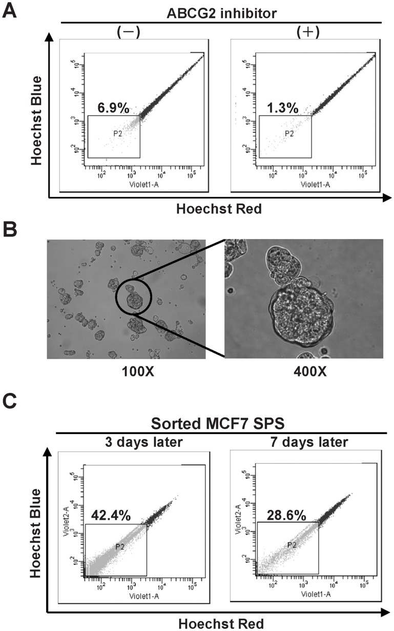
Figure 1. Isolation of breast cancer SPS.
(A) MCF7 cell line exhibiting low Hoechst 33342 staining defines the fraction of SP. The presence of the ABCG2 inhibitor GF120918 blocks the exclusion of Hoechest dye and confirms the selection of SP for further studies. (B) MCF7 SPS generated by SP grown in a serum-free suspension culture (magnification, 100× (left) and 400× (right)). (C) MCF7 SPS repopulated both SP and non-SP cells. The fraction of SP decreased during repopulation. (left on Day 3; right on Day 7).

