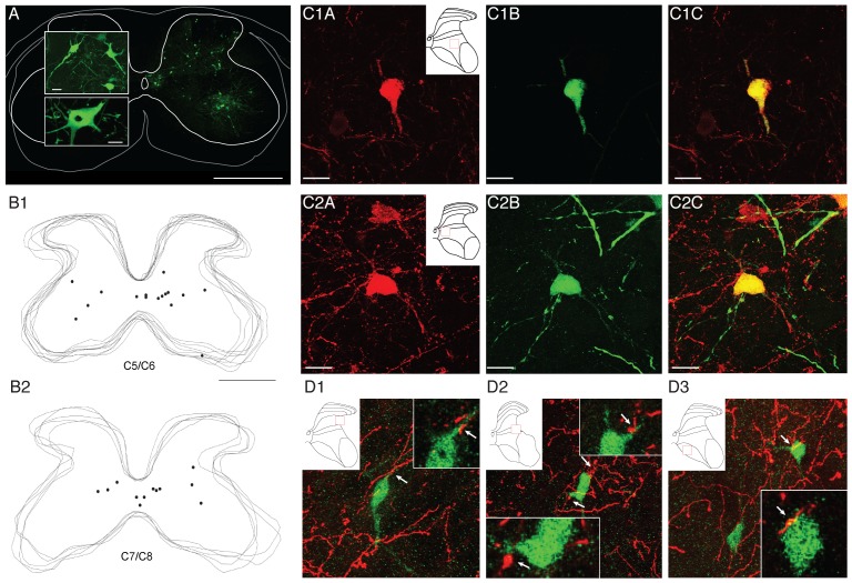
Figure 7. Spinal labeling of last-order (premotor) interneurons with PRV, co-labeling with ChAT, and contacts with CSTs.
(A) Example of labeling in one 40 µm section achieved at 64 hours after intramuscular PRV injection. Large panel shows labeling on ipsilateral side (Calibration: 500 µm). There was minimal contralateral labeling. Top inset shows an example of labeled interneurons (located in lamina 4 on the section shown), and lower panel, a motoneuron, at higher magnification (Calibration: 25 µm). (B1–B2) Overlaid section images for two representative animals processed in Neurolucida showing positions of individual last-order interneurons from PRV injected into the deltoids, biceps and wrist extensors at levels C5/C6 (B1) and C7/C8 (B2) that also label positively for ChAT. (C1–C2) Confocal images of two representative PRV-ChAT double-labeled interneurons at levels C7/C8 (C1) and C5/C6 (C2). ChAT = red; PRV = green. (D1–3) (D) Confocal images of PRV-labeled interneurons receiving contacts from BDA-labeled CST axons terminals. PRV was injected into the biceps and wrist extensor compartments. Each panel shows a projection image (center, large image) and representative 1 µm optical slices (insets). Arrows show sites of contact,. Scale bars for A, large panel = 500 µm, smaller panels = 50 µm; B, same as A; C and D = 20 µm.

