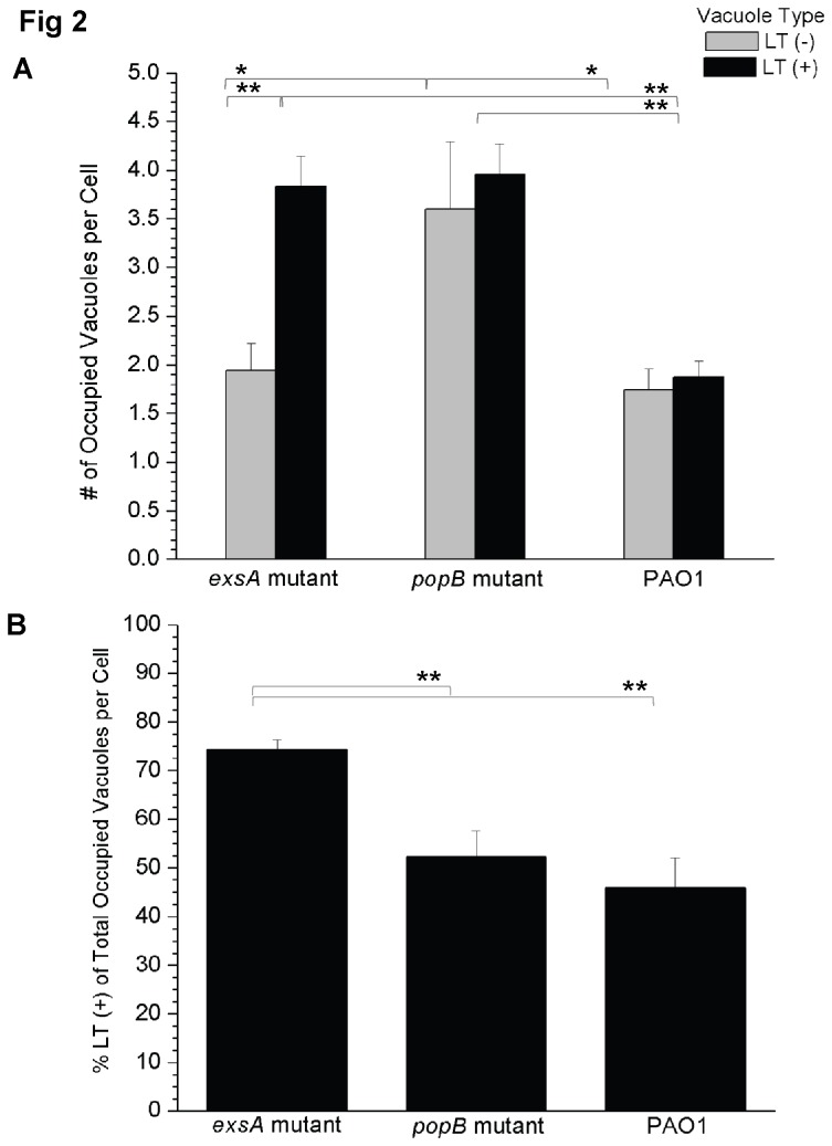
Figure 2. Quantification of acidified versus non-acidified vacuole occupation by wild-type P. aeruginosa and its type III secretion mutants.
(A) Confocal microscopy images were used to classify bacteria-occupied vacuoles in human corneal epithelial cells as either LT (+) (acidified) or LT (-) (non-acidic) at 5 h post-infection with P. aeruginosa PAO1 or its type III secretion mutants (exsA or popB). The data are shown as the mean (+/- SEM) number of bacteria-occupied vacuoles per cell. Grey columns denote LT (-) vacuoles, black columns LT (+) vacuoles. The exsA and popB mutants were both associated with increased numbers of acidified LT (+) bacteria-occupied vacuoles per cell compared to wild-type PAO1 (p < 0.001, Welch’s corrected t-Test). The exsA mutant showed more acidified than non-acidified bacteria-occupied vacuoles per cell (p < 0.001, Welch’s corrected t-Test). (B) To normalize differences in internalization and replication, the percentage of LT (+) bacteria-occupied vacuoles was calculated as a function of the total number of bacteria-occupied vacuoles per cell. Mean percentage (+/- SEM) is shown. The exsA mutant was associated with more acidified bacteria-occupied vacuoles per cell than either the popB mutant or wild-type bacteria (p < 0.001, Welch’s corrected t-Test). A representative experiment of 3 independent experiments is shown in both panels (A) and (B). Calculations excluded cells showing bleb-niche formation. Significant differences between all groups were identified using ANOVA analysis (p < 0.0001), and characterized on a pairwise basis using Welch’s correct t-Test [*p < 0.05, **p < 0.001].

