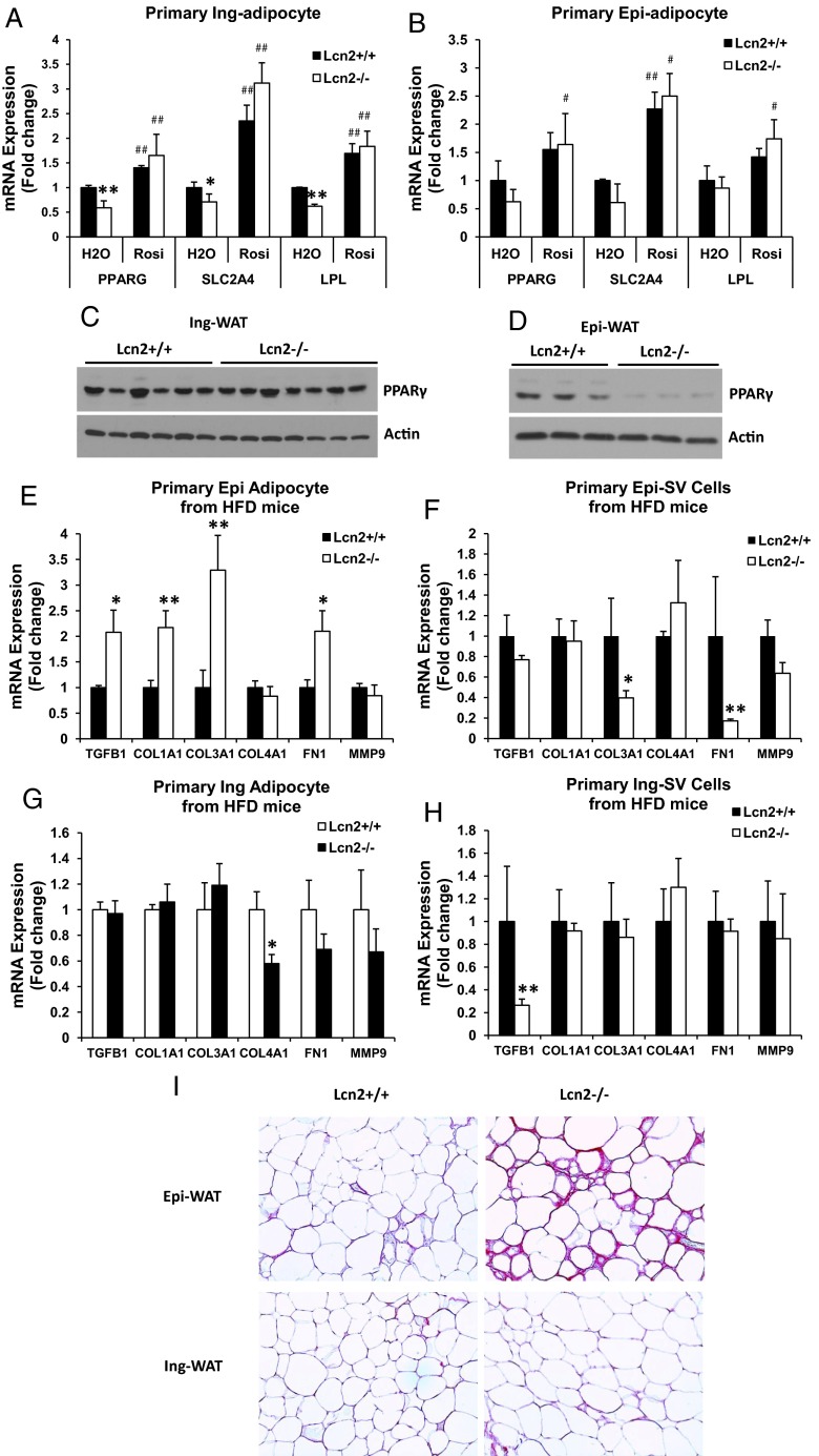
Figure 3.
A and B, The mRNA expression of adipogenic genes in primary inguinal (A) and epididymal (B) adipocytes isolated from WT and Lcn2−/− mice fed an HFD for 16 weeks. C and D, PPARγ protein expression in Epi-WAT (C) and Ing-WAT (D) of HFD-fed WT and Lcn2−/− mice. E–H, The mRNA expression of ECM molecules in epididymal adipocytes (E) and SV cells (F) and inguinal adipocytes (G) and SV cells (H) isolated from WT and Lcn2−/− mice fed an HFD. I, Picrosirius staining of epididymal and inguinal adipose tissue from HFD-fed WT and Lcn2−/− mice; collagen fibers are stained with red. Results (A and B and E–H) represent mean ± SE of 4 to 6 animals. *, P < .05; **, P < .01; WT vs Lcn2−/−; #, P < .05; ##, P < .01, H2O vs Rosi.

