Figure 2.

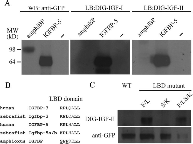
Amphioxus IGFBP Does Not Bind to IGF-I or -II but Gains the IGF-Binding Function with a Single Amino Acid Change. A, Western immunoblot and ligand-blot analysis of amphiBP and human IGFBP-5. Conditioned media from HEK 293T cells transfected with expression plasmids for the indicated IGFBP were collected and analyzed by Western immunoblot (WB) and ligand immunoblot (LB) using the indicated antibody or DIG-labeled IGFs. B, Alignment of the IGF-binding site sequence in the indicated IGFBPs. Bold letters indicate the residues known to be critical for ligand binding. The 2 residues unique to amphioxus and tested are underlined. C, Western immunoblot and ligand-blot analysis of amphioxus IGFBP and the indicated mutants. Conditioned media prepared from HEK 293T cells transfected with expression plasmids for amphiBP or the indicated mutants were analyzed by Western immunoblot using a GFP antibody and ligand blot using DIG-labeled IGF-II. LBD, ligand-binding domain; WT, wild type.
