Figure 2.

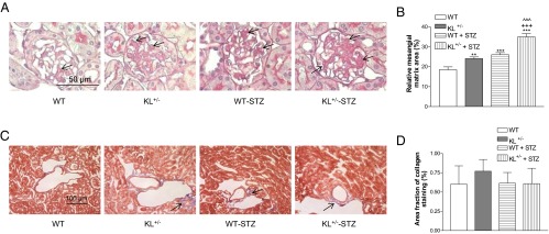
Effects of klotho deficiency on mesangial matrix expansion and collagen deposition in diabetic kidneys. KL+/− mutant and WT male mice were injected with STZ or citrate buffer. Animals were killed 5 weeks after the initial injections. A, Representative photomicrographs of PAS-stained kidney sections. Arrows indicate PAS-positive mesangial material (pink). B, Quantitative measurements of mesangial matrix expansion. Relative mesangial matrix area is expressed as PAS-positive mesangial matrix per total glomerular tuft cross-sectional area. An average value was obtained from analyses of 20 glomeruli per mouse. C, Representative photomicrographs of Masson's trichrome-stained kidney sections. Arrows indicate trichrome-positive collagenous components in cortical interstitium (light blue). D, Quantitative analysis of collagenous components in renal cortex. Images of cortex from 3 consecutive sections for each animal, collected at equal exposure conditions under Nikon Eclipse Ti microscopy, were used in the analysis. Data are shown as mean ± SEM; n = 8. **, P < .01; ***, P < .001 vs the WT group; +++, P < .001 vs the WT-STZ group; ^^^, P < .001 vs the KL+/− group.
