
Fig. 1.
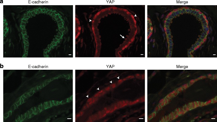
YAP expression in human and mouse normal breast tissue. a YAP expression in normal human breast tissue. Shown are immunofluorescence for YAP (middle panel, red) and E-cadherin (left panel, green). Nuclei were visualized using DAPI. Right panel depicts the merged image. Luminal epithelial cells form clear AJ and are characterized by low cytosolic YAP expression. Note the predominant nuclear YAP localization in myoepithelial cells (arrowheads) and expression of YAP in apical snouts (arrow). Size bar = 5 μm. b. YAP expression in mouse mammary glands. Shown are immunofluorescence for YAP (middle panel, red) and E-cadherin (left panel, green). Arrowheads depict nuclear YAP expression in mouse myoepithelial cells. Nuclei were visualized using DAPI. Right panel depicts the merged image. Size bar = 5 μm