
Fig. 2.
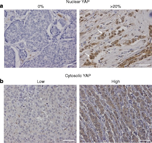
YAP expression in human invasive breast cancer. a Nuclear YAP expression patterns. Shown are representative examples of immunohistochemistry of YAP (IHC). The percentage of nuclei that showed YAP expression was determined and scored as 0 % (left panel) or more than 20 % (right panel). Arrows denote nuclear staining. b Cytosolic intensities of YAP expression. Shown are representative examples of immunohistochemistry of YAP (IHC). Cytosolic YAP expression was scored as either low YAP (left panel) or high YAP (right panel). The sample shown in the right panel was also scored <20 % (15–20 %) for nuclear YAP localization. Size bar = 25 μm