
Fig. 4.
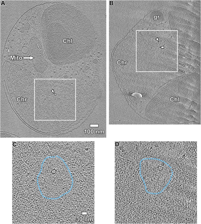
Spindle microtubules reside in a nucleosome-depleted zone. a, b Tomographic slices (10 nm) of two mitotic O. tauri cells, taken at the spindle tunnel. The chromatin (Chr), mitochondrion (Mito), and chloroplast (Chl) are indicated. Each tomogram was rotated to a view along the longitudinal axis of the spindle microtubules (arrowhead), one of which is incomplete (b). As a result of the image rotation, crevasses (arrows) are visible in the right-hand side of b. Subvolumes containing the spindle microtubule(s) and the spindle tunnel are boxed and enlarged in c and d, corresponding to the cells (a) and (b), respectively. The boundary surrounding the nucleosome-depleted zone is delineated by the blue dotted line