
Fig. 5.
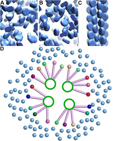
O. tauri chromatin is disorganized. Cartoon models of nucleosomes (blue disks) in O. tauri chromatin in a interphase and b mitosis. In textbook models (c), chromatin is universally presented using the 30-nm fiber; modeled from Scheffer et al. (2011). d Hypothetical model of O. tauri polymer melt chromatin in a mitotic cell, viewed along the spindle axis. Canonical nucleosomes (light blue spheres) and centromeric nucleosomes from nonhomologous chromosomes (multicolored spheres) are positioned around the spindle. Kinetochore protein complexes (lilac rods) connect the centromere to the spindle microtubules (green rings)