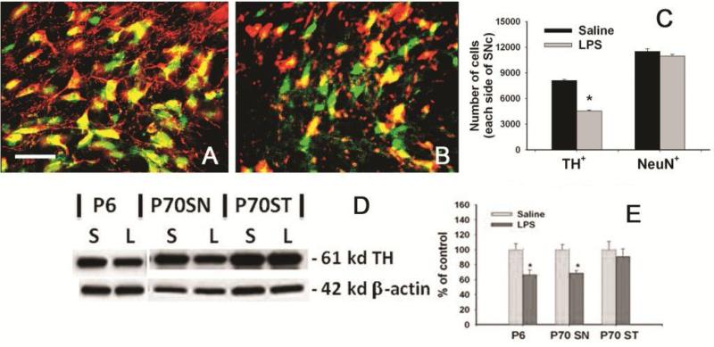
Figure 4.
Suppression of TH expression in neurons from the SN of P70 rats with neonatal LPS exposure. Representative merged photomicrographs of TH+ (red) and NeuN+ (green) cells in the SN of the rat brain from the control group and the LPS-exposed group are shown in A and B, respectively. Stereological cell counting showed that neonatal LPS exposure reduced number of TH+ cells, but not NeuN+ cells in the SN of P70 rats (C), suggesting that neonatal LPS exposure suppressed TH expression, but did not cause significant neuronal death in the SN of P70 rat brain. Western blots for TH expression in the whole brain of P6 rats or in the SN or ST of P70 rats (D) and quantification of the blotting data (E) are consistent with immunohistochemistry results. The results in C and E are expressed as the mean±SEM of 5 animals in each group, and analyzed by t-test. *P<0.05 different from the saline-injected group.

