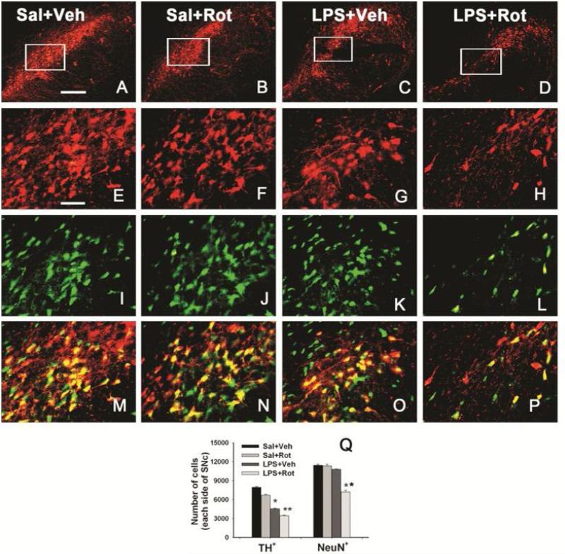
Figure 5.
Neonatal systemic LPS exposure enhanced rotenone effects on the number of TH+ or NeuN+ cells in the SN of P98 rat brain. Representative photomicrographs of TH+ cells (red) at low magnification (A-D) or high magnification (E-H), NeuN+ cells (green, I-L), and the merged images (M-P) in the SN of rat brain are shown here. Representative images for the saline+vehicle group are shown in A, E, I and M; those for the saline+rotenone group in B, F, J and N; those for the LPS+vehicle group in C, G, K and O; and those for the LPS+rotenone groups in D, H, L and P. Neonatal LPS alone reduced the number of TH positive cells (G) in the SN of P98 rat brain, but did not alter the number of NeuN positive cells (K) in the same area. Rotenone treatment in adult life also slightly decreased the number of TH positive cells (F), but not the number of NeuN positive cells (J) in the saline+roptenone rat brain. Rotenone treatment resulted in not only a further decreased number of TH positive cells (H), but also a significant loss of NeuN positive cells (L), indicating actual death of dopaminergic neurons in the SN. The scale bar shown in A (for A-D) represents 200 μm and that in E (for all other images) 50 μm. Stereologic cell counting data of the number of TH+ cells and the number of NeuN+ cells in the SN area are shown in Q. The results are expressed as the mean±SEM of five animals in each group, and analyzed by one-way ANOVA. *P<0.05 different from the Sal+Veh group; ** P<0.05 different from for the LPS+Veh group.

