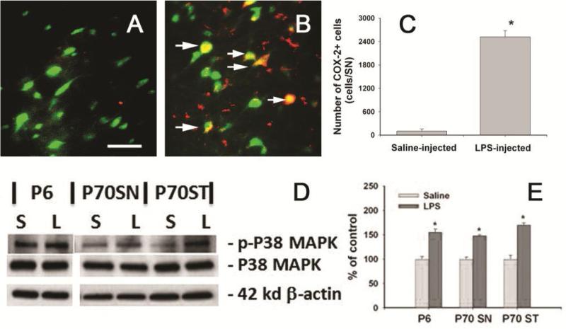
Figure 8.
Neonatal LPS exposed persistently increased COX-2 expression and phosphorylation of P38 MAPK in the P70 rat brain. Representative photomicrographs of COX-2+ (red) and NeuN+ (Green) cells in the SN of P70 control rat brain and LPS-exposed rat brain are shown in A and B. Increased COX-2+ cells were observed in the SN of LPS-exposed rat brain, while almost no COX-2+ cells were found in the control rat brain (A). Many COX-2+ cells found in the LPS-exposed rat brain were NeuN+ neurons (arrows indicated in B). Stereological cell counting data of COX-2+ cells are shown in C. Western blot analysis (D) showed that neonatal LPS significantly increased P38 phosphorylation in the P6 rat brain and the SN and ST of P70 rat brain. Semi-quantification of the blotting data is shown in E. The results in C and E are expressed as the mean±SEM of five animals in each group, and analyzed by t-test. * P<0.05 different from the saline group

