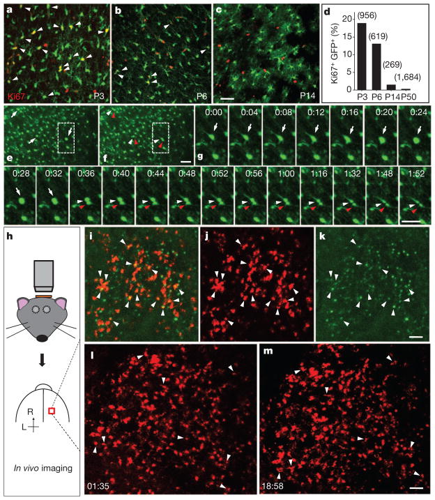
Figure 3. Time-lapse imaging of local proliferation of astrocytes.
a–c, Proliferating astrocytes (arrowheads) in the cortex of hGFAP-GFP transgenic mice, at P3 (a), P6 (b) and P14 (c). d, Summarized data for the percentage of Ki67+GFP+ cells among GFP+ cells with strong GFP signals. e, f, Sequential images of a cortical slice from a P3 hGFAP-GFP transgenic mouse (e, parent cells (arrows); f, daughter cells (arrowheads)). g, Time-lapse images (1 h 52 min) of a dividing GFP+ cell in e and f. h, Procedure to image cell division in vivo. i–k, Images from a P4 triply transgenic hGFAP-CreER;Ai14;CAG-Fucci-Green mouse (i, combined images; j, tdTomato; k, mAG signal; arrowheads, dividing astrocytes). l, m, Time-lapse images at 1 h 35 min (l) and 18 h 58 min (m) from a P5 hGFAP-CreER;Ai14 transgenic mouse (arrowheads, dividing astrocytes). Scale bars, 40 μm (a–g) and 100 μm (k, m).

