
Figure 1.
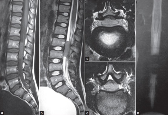
Pre-operative magnetic resonance imaging of the spine showing a sausage shaped intradural extramedullay lesion at L1 and L2 spinal levels. (a) T1-weighted sagittal, (b) T2-weighted sagittal, (c) T1-weighted axial, (d) T2-weighted axial, and (e) MR myelogram