Figure 2.

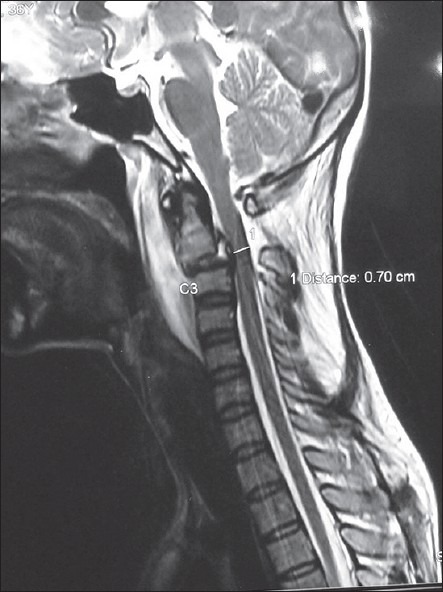
Sagittal T2-weighted magnetic resonance imaging sequences in patient 1 with type IIa Hangman's fracture showing hematoma extending from clivus to C4 along with anterior translation and angulation of C2 over C3

Sagittal T2-weighted magnetic resonance imaging sequences in patient 1 with type IIa Hangman's fracture showing hematoma extending from clivus to C4 along with anterior translation and angulation of C2 over C3