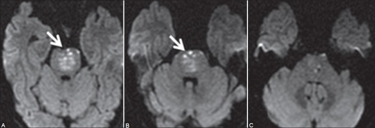
Figure 1 (A-C).

Initial MRI of brain on admission: Axial diffusion-weighted images (A, B) New tiny hyperintense signal foci in ventral and paramedian pons on both sides (arrows) suggestive of acute infarcts. The central paramedian pons reveals lesser hyperintensity due to subacute infarct. At this time, MCPs were normal (C)
