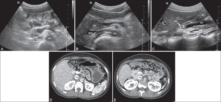
Figure 1 (A-E).

Transverse scans of epigastrium in one of the patients showing absence of body and tail of the pancreas with the fluid distended stomach (ST) seen in the pancreatic bed (A). A slightly caudal section (B) Shows the uncinate process (UP) of the pancreas. (C) Sagittal scan through the superior mesenteric vein (SMV) confirming that the pancreas is not seen superiorly or inferiorly. (D, E) Images of CT scan confirming agenesis of dorsal pancreas (AO: Aorta, IVC: Inferior vena cava, SA: Splenic artery, ST: Stomach, SV: Splenic vein)
