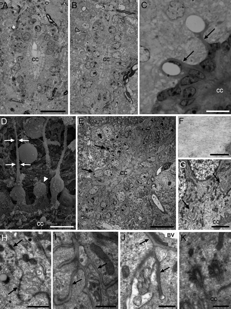
Figure 1. Central canal ependymal (Ecc) cells.
A) Toluidine blue-stained semithin section of the cervical spinal cord, where the central canal shows an oval shape. B) The lumbar central canal is dorsoventrally elongated. C) Central canal cells in the lateral wall, with radial expansions (arrows) in contact with blood vessels. D) Scanning electron microscopy (SEM) image of the canal surface (below). Some cells show long and thin basal processes (arrows), and other cells show rounder cell bodies, without radial process (arrowhead). E) Cells in the central canal are organized as a pseudostratified epithelium, with most nuclei located in the apical row, and some of them in basal position (arrows). The dorsal and ventral regions of the canal (up and below respectively) contain elongated Ecc cells with long radial processes. Blood vessels in close association to the canal can be observed. F) Detail of intermediate filaments of an Ecc cell. G) Horse-shoe shaped Golgi apparatuses polarized with the cis-side pointing at the apical surface (arrows). H) Large junction complexes are found apically between adjacent cells (arrows). I) Basal lamina network flowing through Ecc cells (arrows). J) Radial process of a central canal ependymal cell in contact with the basal lamina (arrows) of a blood vessel. K) Two large and electron-dense basal bodies of a biciliated Ecc cell. A–C are toluidine blue-stained semithin sections, and E–K are transmission electron microscopy (TEM) images. CC, central canal; BV, blood vessel. Scale bar in A (A–B) and E, 20 µm; C and D, 10 µm; F, 200nm; G, 2 µm; H, 1 µm; I, J and K, 500 nm.

