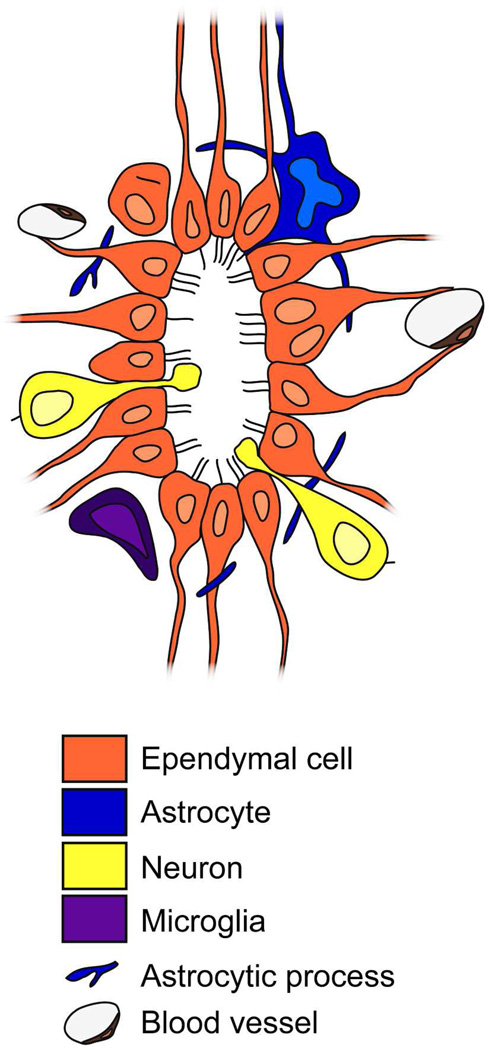
Figure 11. Model for the mouse central canal organization.
Transverse schematic view showing the different cell types found in the central canal. We describe ependymal cells with up to four cilia, most frequently biciliated. Some binucleated cells are present. There are also astrocytes and neurons in contact with the canal lumen. Microglial cells and astrocytic processes surround the epithelium. Blood vessels are very close to the central canal, with an extensive basal lamina network.

