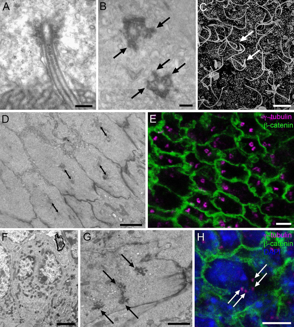
Figure 2. Cilia and basal bodies of Ecc cells.
A) Longitudinal TEM section of a cilium, showing the axoneme with a central pair of microtubules and a large electron-dense basal body. B) Tangential TEM section of the canal surface, showing basal bodies with multiple electron-dense appendages organized as radial spikes (arrows). C) SEM image of the canal surface, were long cilia can be observed as they emerge in parallel (arrows). No multiciliated cells were observed. D) Tangential TEM section of the canal surface, showing that biciliated Ecc cells´ basal bodies are oriented in the same direction and at a similar distance (double sided arrows: 1 µm). E) Immunofluorescence on central canal whole mount preparation. Donut-shaped basal bodies can be observed, stained with γ-tubulin (magenta), and limits between adjacent cells can be followed with β-catenin expression (green). F) TEM image showing an Ecc cell with two distinct nuclei, confirmed by serial ultrathin sectioning. G) En-face TEM section of the central canal surface showing one cell with four basal bodies (arrows). H) Confocal immunostaining on central canal whole mounts confirmed the presence of cells with two nuclei (DAPI, blue), and four basal bodies (γ-tubulin, magenta). Cell contours are labeled in green (β-catenin). Scale bar in A and B, 200 nm; C, 5 µm; D, E, F and H, 2 µm; G, 1 µm.

