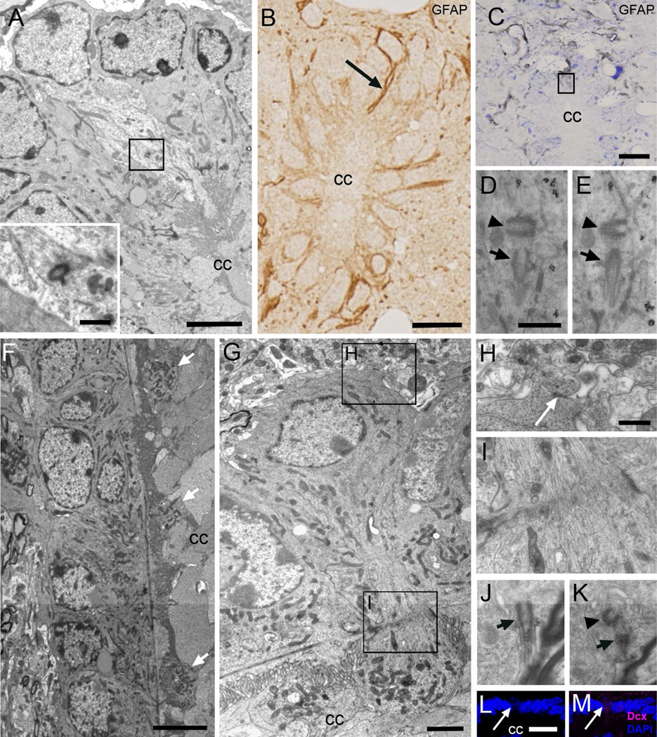
Figure 3. Astrocytes and neurons in contact with the central canal (Acc and Ncc cells).
A) Ultrastructure of an Acc cell with light cytoplasm in contact with the canal in the dorsal region in a transverse TEM section, showing an orthogonally oriented centriole (inset). B) Post-embedding immunocytochemistry on semithin sections, showing a thin GFAP+ expansion in contact with the canal lumen (arrow). C–E) Pre-embedding immunogold (detection of GFAP. C) Toluidine blue-stained semithin section showing grey immunogold-silver labeling for GFAP around the central canal and in thin expansions in contact with its lumen (boxed area). D–E) TEM serial sectioning of the box in C. One of the labeled cells displays a typical single cilium basal body (arrow) with an orthogonally oriented centriole (arrowhead). F) Longitudinal TEM section of the canal where globular expansions of several Ncc cells can be detected (arrows). G) These neurons show a round soma, and their cytoplasm contains abundant mitochondria and RER. H) Ncc cells establish axo-somatic synaptic contacts (arrow) in the surrounding neuropil. I) The apical narrowing contains abundant microtubules. J–K) Single cilium (arrow) with centriole (arrowhead) in a different Ncc cell that was serially reconstructed. L–M) Confocal immunofluorescence shows that Ncc cells express DCX (magenta) and their nuclei are weakly stained with DAPI (blue). CC, central canal. Scale bar in A and G, 2 µm, inbox, 500 nm; B, C and L, 10 µm; D (D–E) and H (H–K), 500 nm and; F, 5 µm.

