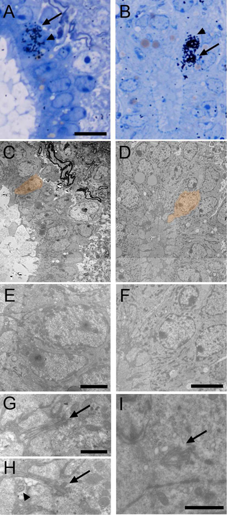
Figure 9. Identification of generated cell types with TEM.
Autoradiography on animals sacrificed 2.5 weeks after [3H]thymidine injections. Labeled cells were identified on toluidine blue-stained semithin sections (A-B, arrows), serial ultrathin sectioned and analyzed under the TEM (C–I). Pairs of labeled cells were frequently observed (arrows and arrowheads). Electronmicrographs of cells pointed with an arrow and depicted in orange are shown. A) Labeled Ecc cell (C and E) with two cilia, appearing on two different ultrathin sections (G–H). These cilia showed 9+2 structure (arrowhead) and large basal bodies (arrows). B) Labeled uniciliated Ecc cell (D and F) showing the beginning of the cilium with a large basal body (I, arrow). Scale bar in A (A–D), 10 µm; E, 2 µm; F, 5 µm; G (G–H) and I, 1 µm.

