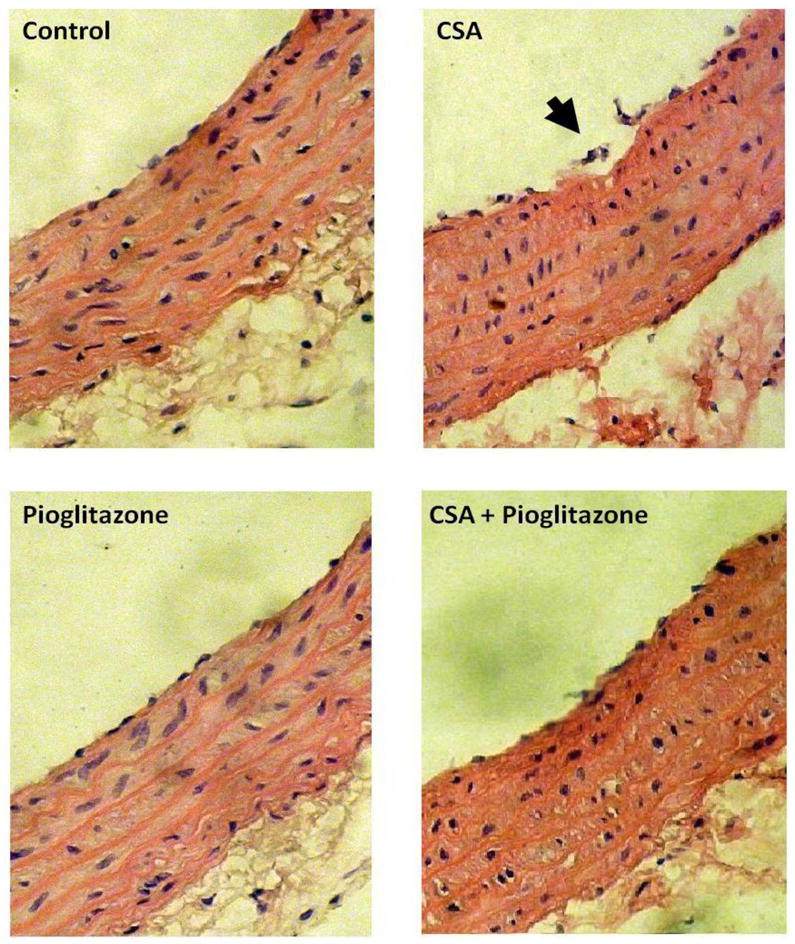
Figure 8.

Hematoxylin and Eosin stained photomicrographs (× 100) of aortas obtained from rats treated with cyclosporine (CSA, 20 mg/kg/day), pioglitazone (2.5 mg/kg/day) or their combination for 14 days. In contrast to the intact endothelial cell layer seen in vehicle (olive oil) preparations, the aorta of CSA-treated rats shows focal disruption of the endothelial lining (arrow). This effect of CSA disappears in preparations treated concurrently with pioglitazone.
